Sars Cov -2.jpg
Sars Cov-2 thumbnail.jpg

Sars-Cov-2

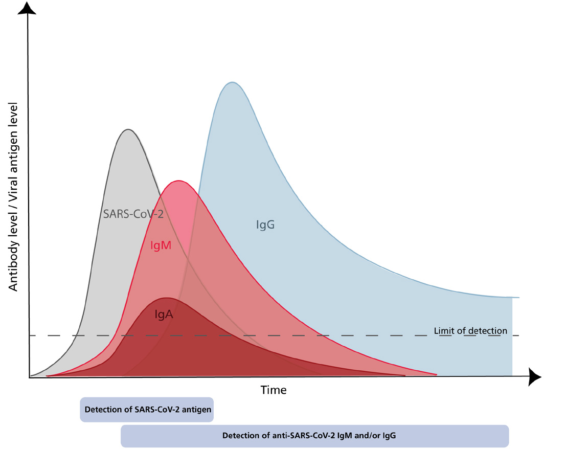
The Global emergence of SARS CoV-2

In December 2019, a new severe respiratory disease emerged in the province of Wuhan, China. Later, a novel coronavirus (CoV) named Severe Acute Respiratory Syndrome Coronavirus 2 (SARS-CoV-2; formerly 2019-nCoV) was identified as the aetiological agent of that disease (COVID-19) (Lake 2020; Gorbalenya et al. 2020).

SARS-CoV-2 shares 79% homology with the severe acute respiratory syndrome coronavirus (SARS-CoV) described in 2002/2003 (Lu et al. 2020; Zhou et al. 2020). Both viruses belong to the family of Coronaviridae which are enveloped, positive-sense, single-stranded RNA viruses that can occur in various animal species. Most CoVs affecting humans are causing mild respiratory infections, only the Middle East respiratory syndrome (MERS)-CoV had been reported to be responsible for severe courses of disease besides the SARS viruses.

SARS-CoV and SARS-CoV-2 both use their transmembrane spike (S) glycoprotein to bind the hACE-2 receptor on host cells for cell entry (Tortorici and Veesler, 2019; Zhou et al. 2020).

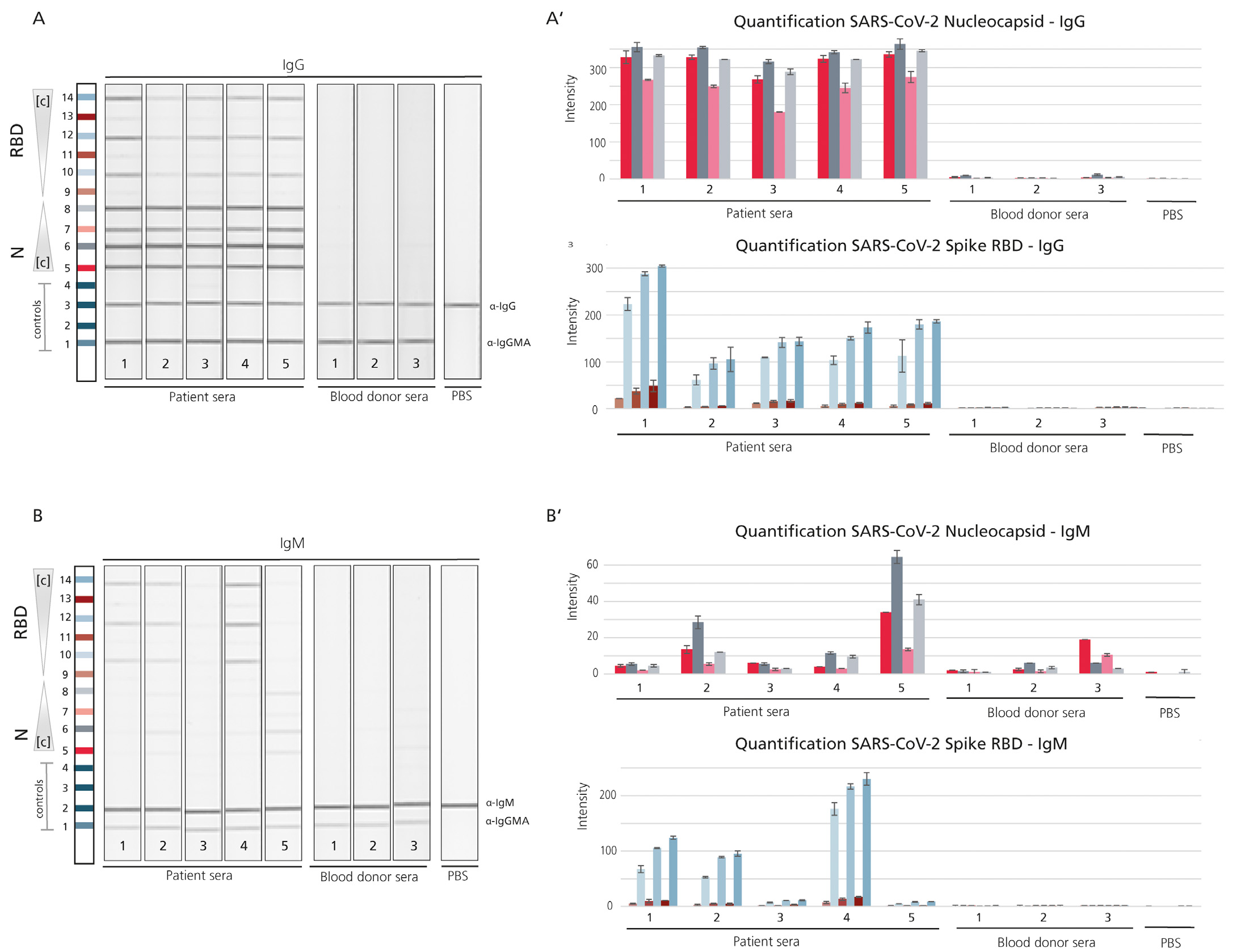
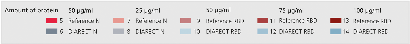
The S glycoprotein is composed of two functional subunits and the S1 subunit mediates binding to the host cell. The S protein is very immunogenic and, as it is mediating binding to the host cell receptor, neutralizing antibodies often target its receptor binding (RBD) domain (Amanat et al. 2020). Sequencing revealed that the receptor-binding spike protein encoded by the S gene was highly divergent from other CoVs, with less than 75% nucleotide sequence identity to all previously described SARS-CoVs, except for a 93.1% nucleotide identity to RaTG13, the closest ancestor of the virus occurring in bats (Zhou et al. 2020).

The protein sequence of the S gene of SARS-CoV-2 shows three short insertions in the N-terminal domain as well as changes in most of the key residues in the RBD compared with the sequence of SARS-CoV (Zhou et al. 2020). This highly diverged motif might be a good target for serological assays that aim to distinguish between the antibody responses against the two SARS virus strains as well as the CoVs causing common cold.

The nucleocapsid (N) protein forms complexes with genomic RNA, interacts with the viral membrane protein and seems to play a critical role in enhancing the efficiency of virus transcription and assembly (McBride et al. 2014). Like the S protein, the N protein is highly immunogenic as shown earlier in studies on SARS-CoV (Qiu et al. 2005).

Sars-CoV-2 Nucleocapsid (N) Protein, SARS-CoV-2 Spike (S) Glycoprotein, SARS Antigens Ordering Information

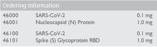
Please contact our Customer Services Team directly to discuss your requirements on tel:+44 (0) 1495 363000 or alternatively you can submit an enquiry here.

Technical information

In a typical immunological response IgM antibodies are usually the first line of defence. Seroconversion to IgG then occurs later on and these high-affinity IgG antibodies will be responsible long term immunity and immunological memory (Li et al. 2020; Racine et al. 2009).


Figure 1: Schematic representation of typical adaptive immune response after viral infection during course of disease and convalescence.

Due to the late onset of antibody development during the course of the disease, molecular methods will be preferred for diagnostics of SARS-CoV-2. However, sensitivity limitations were observed in molecular testing and serology could aid to identify patients appearing false-negative in PCR despite showing symptoms (Farnsworth and Anderson 2020; Xu et al. 2020). Further, serology is strongly suggested to play a major role for screening populations to determine exposure as well as potential immunity, to identify convalescent individuals as plasma donors as well as for research on immune response and to help identify neutralizing antibodies (Amanat et al. 2020; Farnsworth and Anderson 2020; Okba et al. 2020).

A prerequisite of developing highly sensitive serological assays for diagnostic purposes is the use of high quality raw materials that reliably enable distinguishing healthy individuals from those infected. One of the core raw materials of such tests are antigens capable of capturing the patient’s antibodies of interest. DIARECT has newly developed SARS-CoV-2 Nucleocapsid (N) Protein and SARS-CoV-2 Spike (S) Glycoprotein outperforming reference antigens  (manufactured in different expression systems) in different serological assays. DIARECT’s antigens showed a higher reactivity towards patient serum with comparably low background in negative samples at the same time (Figure 2).


  Figure 2: Comparison of DIARECT‘s Nucleocapsid (N) antigen and Spike (S) RBD antigen to respective reference products.


Figure 2: Comparison of DIARECT‘s Nucleocapsid (N) antigen and Spike (S) RBD antigen to respective reference products

A; B: Line Assay. The antigens and controls (IgG; IgM; IgGMA) were printed on a nitrocellulose membrane as indicated. Antigens were printed at the following concentrations: Line 5 (Reference N) 50 µg/ml; Line 6 (DIARECT N) 50 µg/ml; Line 7 (Reference N) 25 µg/ml; Line 8 (DIARECT N) 25 µg/ml; Line 9 (Reference RBD) 50 µg/ml; Line 10 (DIARECT RBD) 50 µg/ml; Line 11 (Reference RBD) 75 µg/ml; Line 12 (DIARECT RBD) 75 µg/ml; Line 13 (Reference RBD) 100 µg/ml; Line 14 (DIARECT RBD) 100 µg/ml. Each line assay was incubated with a patient serum (1-5), serum from a healthy blood donor (1-3) or PBS (negative control). Detection was made using anti-human IgG (Figure 2 A) or anti-human IgM (Figure 2 B) secondary antibodies. Each line was prepared in duplicates (only one shown per serum).

A‘; B‘: Graphic evaluation of the line assays. Mean of intensities of line assays (n=2) including standard deviations are displayed.

DIARECT’s SARS-CoV-2 antigens are produced in the baculovirus/insect cell expression system.

References:

Amanat et al. (2020) Nat Med. 26 (7): 1033 - 1036
Farnsworth and Anderson (2020) Clin Chem. hvaa107
Gorbalenya et al. (2020) Nat Microbiol. 5 (4): 536 - 544
Lake (2020) Clin Med (Lond). 20 (2):124 - 127
Li et al. (2020) J Med Virol. 92: 1518 - 1524
Lu et al. (2020) Lancet. 395 (10224): 565 - 574
McBride et al. (2014) Viruses. 6 (8): 2991 - 3018
Okba et al. (2020) Emerg Infect Dis. 26 (7)
Racine et al. (2009) Immunology Letters 125 (2): 79 - 85
Qiu et al. (2005) Microbes Infect. 7 (5-6): 882 - 889
Tortorici and Veesler (2019) Adv Virus Res. 105: 93 - 116
Xu et al. (2020) Emerg Microbes Infect. 9 (1): 924 - 927
Zhou et al. (2020) Nature. 579 (7798): 270 - 273

NB - In some countries the use of certain antigens in diagnostic tests may be protected by patents. DIARECT is not responsible for the determination of these issues and suggests clarification prior to use.

Technical Documents

If you require any further product specific technical information or datasheets please submit your request to our technical team here 

Last updated: 22/03/2022

SARS CoV 2 Antigens Brochure

There are currently no documents in this category.

Request a callback

Newsletter

Receive regular industry insights and the latest BBI Solutions' news & product launch information by signing up to our Newsletter